by Dr. David Sabow and Robert J. O’Dowd
Convincing evidence in a scientific paper by two forensic experts of the murder of Colonel James E. Sabow rejected by leading forensic journals.
(RAPID CITY, SD) – The difference between homicide and suicide can sometimes be a close call, requiring extensive pathological review and crime scene reconstruction. However, the violent death of Marine Colonel James E. Sabow at MCAS El Toro on January 22, 1991, was a clear case of homicide. For those with knowledge of this cold case, this isn’t even a close call.
 An NCIS cold case investigation of Colonel Sabow’s death was conducted in September-October 2011. Special Agent Julie Haney, an NCIS cold case specialist, interviewed Dr. Singhania, the Orange County Medical Examiner who conducted the autopsy in January 1991. Dr. Werner Spitz, an internationally recognized pathologist, reviewed the autopsy report and other records at Special Agent Haney’s request.
Dr. Spitz provided a sworn affidavit to Haney stating that Colonel Sabow was murdered and the crime scene was tampered with. A few days later Dr. Spitz orally withdrew his affidavit
Bryan Burnett, a crime scene reconstructionist and friend of Dr. Sabow with expert knowledge of the case, accompanied Haney to the meeting with Dr. Singhania but was left cooling his heels outside the conference room.
There is no transcript of the meeting between Special Agent Haney and Dr. Singhania. And, just to make matters more interesting, Special Agent Haney was part of the original crime scene investigation where there are allegations of crime scene tampering.
The NCIS cold case investigation found no basis for homicide, despite a sworn affidavit in September 2010 from Dr. Werner Spitz, an internationally known pathologist, who reported: the crime scene was tampered with, considerable discrepancies between the written reports and the photographs, considerable doubt about the Coroner’s certification of death as suicide, the certification of suicide was a rush to judgment based on erroneous assumptions, and that Colonel Sabow was murdered.
Special Agent Haney refuses to pursue the case with the U.S. Attorney for lack of lack of proof that a homicide was committed.
If you think there’s more to the NCIS crime scene investigation, you are not alone. In my old neighborhood in Philly, some might even say, ‘the fix is in’.
A recent paper submitted to two forensic science journals by Dr. Sabow and Bryan Burnett provides convincing evidence of homicide and provides support for a rush to judgment by the Orange County Medical Examiner.
However, The Investigative Science Journal editor or the alleged reviewers (the editor never released the reviewers’ comments despite twice being requested for them) did not take the time to read the paper while the Journal of Forensic Science’s reviewers who “apparently had not read the manuscript came up with a criticism that was outrageous,” which resulted in Bryan Burnett filing a complaint with the Ethics Committee of the American Academy of Forensic Sciences (AAFS).
Brian Burnett said that “the chairman of the Ethics Committee just before the AAFS meeting last year apparently decided on his own that the committee did not have jurisdiction in the matter. There was no comment on the issues presented by the reviewer’s comments.”
The only explanation for rejection by the Investigative Science Journal was the ‘bias’ of Dr. Sabow for supporting homicide and not suicide as the manner of death, according to Bryan Burnett.
Rather than judge the paper on its merits, “the reviewers selected by both journals prejudged the submissions with no indication of even reading the submissions. Both editors went along with these outrageous reviewer critiques,” said Burnett. Even a non-scientist layperson knows that this in inherently wrong. What were they afraid of? Or as suggested by the authors, did someone buy off the editors with money or a threat that convinced them to reject the scientific papers outright?
What would the motive be for such an action? Dr. Sabow and others are convinced that an investigation by the U.S. Attorney or an independent council would open a Pandora box to the black operations and narcotrafficing involved in Iran/Contra and possibly reach many key players who are still alive today.
If this murder ever gets to a Federal Grand Jury, look for quick indictments and convictions of those involved in the murder and the conspiracy to commit murder. Nearly 22 years later this is a very big ‘IF’.
You can decide for yourself whether there’s enough evidence to support homicide and the cause for an independent review of the Orange County Coroner office and the Colonel Sabow death certificate by the California Attorney General.
Dr. Sabow’s analysis of the pathology of the murder is provided below in Part I. Bryan Burnett’s detailed Crime Scene Reconstruction analysis is published in Parts II and III.
PATHOLOGY OF THE MURDER
The case presented in this paper will illustrate two principles; the importance of understanding the concept of “sudden” death, in contrast to “instantaneous” death, as well as the necessity of withholding the determination of the “manner of death” in equivocal (accidental, homicide or suicide) death analyses until the totality of evidence, including a psychological autopsy, is available. When the manner of death is “equivocal,” the medical examiner should employ the term “pending” on the death certificate until a comprehensive review of the death investigation has been completed that goes well beyond the autopsy table (Carter, 2006).
The importance in distinguishing between suicide and homicide in death investigations cannot be overstated. The impact on the family of the victim from an emotional as well as a financial standpoint frequently depends on this determination. However, not infrequently, medical examiners do not have the necessary understanding of complex neural systems that must be employed to ensure an accurate determination. A thorough knowledge of the neurophysiology of the brain and brainstem may be required in this process, because the presence or absence of certain elementary brainstem functions and the timing of their loss is pivotal in reaching the correct conclusion. This is an area of study that eludes even the most experienced professionals as is evident in the controversies regarding the subject of death and dying (Abram et al., 1981).
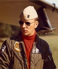
On January 22, 1991 a fifty-one year old Marine Corps colonel was found dead in the backyard of his base home on the Marine Corps Air Station at El Toro, CA (Fig. 1). The colonel, with nearly 30 years of service, was Chief of Operations for Marine Air, Western Area and Assistant Chief of Staff. He was a veteran jet fighter pilot with 221 combat missions in Viet Nam where he received the Bronze Star with Combat “V” for Valor. The colonel was a positive and well-adjusted officer with outstanding “Fitness Reports” throughout his career (United States Marine Corps. Fitness Reports, 1987, 1988, 1989 and 1990). His personal wealth was substantial and his retirement benefits after 30 yrs of service would be sizeable. Overall, his background and circumstances would be the antithesis of a “suicidal proclivity” (Cavanagh, et al., 2003). Several days before his death, he was temporarily suspended from his duties, pending an investigation for alleged improper use of government aircraft. However, only hours before his death, he informed his friend, General J.K. Davis (Retired Assistant Commandant of Marine Corps) of his innocence and that unless all investigations of him were terminated, he would demand a Court Marshall and reveal all of what he knew of irregular flight activity at the base (personal communication to D. Sabow, 1991). These events suggest a motive for the colonel’s homicide.
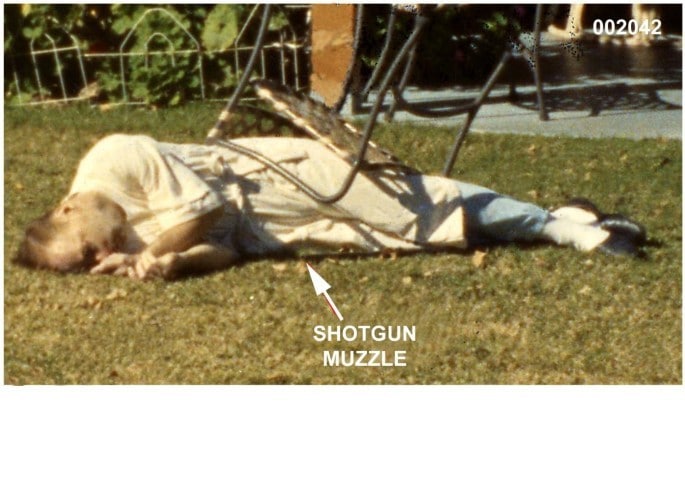
He was found at 0930 by his wife lying on his right side, dressed in a white terrycloth bathrobe, which covered his tee shirt and pajama bottoms, just as he was dressed when she had left him to attend a brief church service at 0830. The victim’s Ithaca double barrel 12 gauge shotgun was in front of him with the stock under his legs and feet. In the crime scene photos a patio chair was on top of him (Fig. 1).
Figure 1. The body in the backyard of his home; the shotgun is in front of him with his legs over its stock. A patio chair is on top of the body.
The decedent’s body was taken to the Orange County Sheriff/Coroner’s Office (California) for the post mortem examination that was performed the following day. There was extensive photographic documentation of both the death scene and autopsy. Fingerprint exemplars and gunshot residue (GSR) samples were obtained at the crime scene and x-rays were taken during the autopsy.
The Medical Examiner completed the death certificate on the same day the autopsy was performed (Singhania, 1991). The “cause of death” was determined to be “massive cerebral contusions and lacerations of the brain secondary to gunshot wound to the head”. The “manner of death” was recorded as “suicide.” It also affirmed “death was immediate.” This was defined on the certificate as “the interval between onset and death.” The “onset” was defined as the “gunshot wound to the head. The brother of the colonel is a board certified neurologist who also has extensive experience (37 years at this writing) as a practicing forensic neurologist and has participated in multiple death investigations and trial in Federal Court. After studying the report and reviewing other information released by the NIS, he had major concerns regarding the “manner of death” determination. His concerns triggered two separate congressional investigations: United States Congress, House of Representatives. Defense Authorization Bill. 1994; Sec. 1185. 1993 and Defense Authorization Bill. 2004; H1588, Division A, Title I, Subtitle I, Section 584. In addition, the Chairman of the House Armed Services Committee, Duncan Hunter (R-CA), demanded a review of the matter in a letter to United States Attorney General Alberto Gonzales in 2007 (Hunter, 2007). The Justice Department declined; denying jurisdiction in the matter (Herting, 2007).
Figure 2. A. Reenactment of the suicide scenario where the victim sat in a chair and inserted the shotgun into his mouth; the muzzle is gripped by the left hand while the right hand reaches down to depress the trigger. The shotgun was found close to the body, partly under the right leg (Fig. 1). This would place the shotgun in the reenactment on the outside of the right leg. In this scenario, blowback from the mouth would spray the thighs (arrows) with blood and tissue. B. Reenactment of part of the homicide scenario where the assailant inserted the shotgun into the mouth of the victim after the blunt force trauma to the back of his head. Arrows: direction of the blowback when the shotgun muzzle is in the mouth at discharge.
If the victim committed suicide and whose suicide comported to the body and shotgun as depicted in the crime scene photos, then the following would have had to occur:
The victim sat in a patio chair while holding a 12-gauge shotgun in his mouth with his left hand. He pushed the trigger of the shotgun with a finger or thumb of his right hand. Then he fell to the right landing on top of the shotgun. The position of the body and shotgun indicates that the stock of the shotgun had to have been on the ground adjacent to the lateral side of his right ankle (Fig. 2A and Anon., 1991, p. 2). The chair toppled on top of him.
Alternatively, if death was a homicide, what follows would most likely had occurred:
The victim received a blow to the back of his head, which caused a depressed skull fracture and a fatal brain injury due to the extensive involvement of the brainstem. Death was not instantaneous following the severe head trauma. Rather, the victim was rendered unconscious and he would have exhibited the typical brainstem signs that forecast impending death. Minutes later and probably after the victim had already expired, an assailant shoved a shotgun into the victim’s mouth and discharged the left barrel (Fig. 2B). The shotgun was then placed in front and partially under the victim. Then a patio chair was placed on top of the victim to complete the suicide appearance (the timing of the placement of the patio chair on the victim is controversial).
THE AUTOPSY
The autopsy was performed at the Orange County Sheriff/Coroner’s Medical Examiner’s facility (California) on January 23, 1991. The following observations were taken directly from the autopsy report (Singhania, 1991) and autopsy photos:
Gunshot wound (entry type) in roof of mouth. No exit wound. Contact wound to soft palate.
Large amount of aspirated blood in the right lung.
Left lung weighs 440 gr., the right weighs 970 gr. [normal adult male is 400 to 450 gr. (Moore, 2009)].
Section of the lung tissue shows congested, atelectactic, edematous, hemorrhagic lung with extrusion of hemorrhagic frothy fluid. (There was no notation of which or whether both lungs were sectioned.)
Pharynx shows a superficial laceration of the right side of the pharyngeal wall.
All brain tissue is massively lacerated including brainstem.
No intact brainstem could be identified including medulla, cerebellar tissue, midbrain, pons, cerebral peduncles. [A remnant of medulla appeared in the foramen magnum at the base of the skull that was contiguous with spinal cord. Upper portion of spinal cord shows evidence of disintegration (which then had to have included the “remnant” of the brainstem seen in the foramen magnum)].
Figure 3. A. The back of the head photographed at the Orange County coroner’s facility prior to autopsy; the swollen area on the back of the head and neck has a patterned ecchymosis (bruising) which reflects the outline of the club used to inflict the injury. The posterior of the right pinna (ear) not covered by blood shows ecchymosis typical of “Battle’s sign” (see text). Note the relative positions of the external ears to the skull: left up against the skull and the right displaced away from the skull by swelling that occurred prior to the victim’s death.B. X-ray of the victim’s skull from the right side showing a depressed right occipital fracture; arrow points to the depressed fracture of the occipital region of the skull behind the right pinna. The small bright objects are shotgun pellets. X-ray image globally enhanced by Adobe Photoshop® levels. C. Autopsy image with the victim’s reflected scalp revealing part of the hematoma that had formed over the depressed skull fracture.
Ecchymosis behind right pinna with massive swelling and displacement of right pinna from skull; contrasting with normal appearing left pinna (Fig. 3A).
Ecchymosis around both eyes (Fig. 5C).
Superficial contusions and lacerations of the lips. Laceration of the anterior [ventral] surface of the tongue and mid tongue area.
Skull X-rays taken at the autopsy show a large depressed occipital skull fracture (3B). On the right side there are subcutaneous and subgaleal hematomas over the depressed occipital skull fracture and “massive scalp hemorrhaging on its inner surface [observed] after the scalp is reflected from posterior to anterior” (Figs. 3A and 4A). In addition, there is massive hemorrhaging of the right temporalis muscle. This contrasts with the left side that has no hematoma or hemorrhage (Fig. 4B) or depression of any skull fragments (Anon., 1991, p. 9).
Figure 4. A. Right scalp reflected from posterior to anterior showing hemorrhage and hematomas over the skull and massive hemorrhaging of under surface of scalp and temporalis muscle. B. Reflection of left scalp shows no hemorrhaging.
DISCUSSION
In equivocal death investigations the medical examiner must always be aware that the autopsy does not determine the manner of death. The autopsy is but one factor to be considered in establishing the manner of death. This classification of the death should reflect the totality of the information that has been gathered in the entire death investigation, which includes the autopsy findings, police and crime scene report, medical records, witness statements, fingerprint results, GSR and blood spatter analysis, social background (psychological autopsy), and any other available reliable data. It should be emphasized that evidence necessary to support a suicide theory should depend upon the unconditional lack of evidence indicative of homicide (Carter, 2006). Since the entirety of evidence is almost never available when completing the death certificate, the “manner of death” on the death certificate should be designated as “pending.”
Depressed Cranial Skull Fracture. From the autopsy we learned that a 2.5 cm diameter, depressed occipital skull fracture was present (Fig. 3B). A photograph taken prior to the autopsy (Fig. 3A) demonstrates massive swelling of the right posterior head and neck. A hematoma was discovered immediately under that swollen area (Anon., 1991, p. 9; Figs. 3C and 4A) and immediately over the depressed skull fracture. All the shotgun pellets were located within the confines of the skull. There were no bone fragments or shotgun pellets within the hematoma. The x-ray (Fig. 3B) shows the fractured bone protruding inwardly not outwardly as would occur if the fracture was the result of the intraoral shotgun wound. Furthermore, since the victim was right handed, the pellets should have been directed from right to left and any localized shattering of the skull should have been on the left side.
Figure 4 demonstrates the contrasting appearances between the two sides when the scalp is reflected from anterior to posterior. Four to five gallons of hot explosive gases are generated within the skull with an intraoral shotgun wound (Di Maio, 1999). This causes a symmetrical distending force against the inner skull surface and the resulting injuries should also have been symmetrical. Indeed, the skull x-rays show irregular long fracture lines on both sides where the skull bones have separated from the explosive force within. In this case, the skull, the scalp and the hemorrhaging were entirely different on the two sides. An evaluation of the skull x-rays and correlating them with the soft tissue damage observed on the right side of the skull, points to blunt force injury to the right back of the head before the colonel received the intraoral shotgun wound.
Figure 5. A. Ecchymosis of the right pinna and a small area anterior to the pinna. B. The left pinna has normal coloration. C. Face of the victim showing ecchymosis of the eyes or “raccoon eyes” typical of basilar skull fracture. There is extensive swelling on the right side of the face.
Basilar Skull Fracture. There is evidence of sustained bleeding from basilar skull (bones at the skull base) fractures in addition to the depressed occipital skull fracture of the cranial vault. A clinical indicator of the presence of a basilar skull fracture is a purplish discoloration of the skin (ecchymosis) of the external pinna and the skin over the mastoid (Fig. 5A) as well as portions of the anterior aspect of the right pinna (Fig. 5A); compare with the normal coloration of the left pinna, Fig. 5B). This is characteristic of a basilar skull fracture of the temporal pyramids at the skull base and is known as Battle’s sign (May, 1977). Figure 5C shows another classic clinical sign of a basilar skull fracture, ecchymosis around the eyes, known as raccoon eyes (Herbella, et al., 2001). This fracture is more anterior and involves the sphenoid bone (May, 1977; Herbella, et al., 2001). Since the tissues at the skull base are firmly adherent, the perfusion pressure must be high enough for the blood to dissect between the bone and its covering tissue to produce these visible subcutaneous hematomas (Wyngaarden, 1985). The presence of these signs indicates the basilar fractures occurred while the victim maintained a significant systolic blood pressure. They are ante mortem signs. If the victim’s death was instantaneous, these classic signs of basilar skull fractures could not have developed. Furthermore, if these classic signs were the result of the boney destruction of the skull base from the shotgun blast, they would have been symmetric and roughly the same on both sides.
Respiratory System. The autopsy report states the victim not only had blood in his large breathing passages, but the blood filled his alveoli, producing hemorrhagic frothy fluid. This was discovered when the lung tissue was sectioned at autopsy. The right lung weighed 970 grams and the left lung 440 grams. The average normal male adult lung weights are 450g (R) and 375g (L) (Moore, 2009). There was an excess of approximately 545g in the right lung. The autopsy report concluded that the victim aspirated almost one-half liter of blood into his right lung. The left lung weighed 440 grams indicating there was very little aspirated blood. The excess right lung weight could not be a result of terminal neurogenic pulmonary edema, because this type of edema would be distributed to both lungs (Baumann, et al., 2007). Likewise, lividity must be ruled out for a similar reason. Lividity is simply a function of the dependant position. The lungs just as the kidneys are “paired” organs. One of each occupies positions on opposite sides of the body. In the abdomen the kidneys are only remotely connected by its main blood vessel, the aorta. The lungs in two sides of the chest have the trachea as there only connection. Blood in the left lung could have accumulated in the medial portion of the left lung and blood in the right lung would accumulate in the lateral portion of that lung due to lividity. But the left lung could not transfer blood to the right lung to account for any extra weight (Standring, 2005).
The approximately one-half liter of blood extra weight of the right lung had to have come from somewhere other than the lungs. The autopsy showed the only lacerated areas of the body that had direct continuity with the right lung were the lips, tongue and pharynx. Therefore they had to be the origins of the blood. Since the autopsy also showed that all the breathing passages were filled with blood-from the trachea, through the bronchi and bronchioles and into the alveoli (Singhania, 1991), then it follows that the origin of that blood was also from those same regions.
The victim was found lying on his right side. And as is seen in Fig. 1, the head is drooped to the ground so that any passive flow from those lacerated areas would have been out through the mouth. Furthermore, even if the passive flow of blood due to gravity is claimed to explain the blood in the large breathing passages, it could not account for the blood filled alveoli. The alveoli contain collagen and elastic fibers. The elastic fibers allow the alveoli to stretch as they fill with air when breathing in. They then spring back during breathing out in order to expel the carbon dioxide-rich air. (Weibel, 1963). The alveoli remain closed until inspiration occurs. Therefore, the gravitational flow of blood could not have entered the alveoli and could not account for the blood filled right lung unless the victim was actively breathing.
Consequently, the victim had to have been lying on his right side when he aspirated the blood into his right lung. Central neurogenic hyperventilation is a characteristic respiratory pattern seen in severe brain stem injuries where the victim is near death (Posner, et al., 2007). After a severe brainstem injury where a laceration of the pharynx was present, the ensuing hyperventilation would explain the presence of one-half liter of blood in the right lung. Furthermore, this breathing pattern continued for a few minutes which accounts for the volume of aspirated blood.

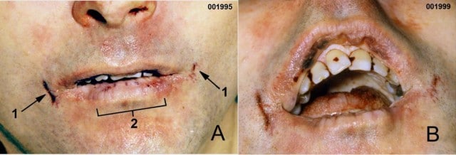
Figure 6. Evidence of the victim biting his lips, likely in decorticate/decerebrate rigidity before the shotgun blast. A. Frontal view, 1: linear abrasions at the angles of the mouth resulted from the stretch of facial tissues at moment of intraoral shotgun discharge. 2: The linear bruising and abrasions coincide with the upper middle and lateral incisors. B. A more inferior aspect image that better shows the swelling of the lower lip, and bruising/laceration of the upper lip by the lower front teeth. Images globally enhanced in Photoshop.
Figure 7. Autopsy images of the tongue. A. Dorsal tongue. B. Ventral tongue; arrows point to bruising and lacerations made by the victim’s teeth.
Oral Mucous Membranes and Tongue. The autopsy photos document lacerations of both the upper and lower lips (Fig. 6) corresponding to teeth injuries, as well lacerations of the anterior [ventral] surface of the tongue and mid tongue area (Fig. 7). These lacerations correspond mostly to the upper and lower incisors and are frequently seen after convulsive epileptic seizures (Brown, et al., 2004). The vertical tears at both sides of the mouth are caused by rapidly expanding gas within the mouth from the shotgun blast (DiMaio, 1999).
Severe brainstem injuries resulting in coma can cause muscular reflex spasms termed decerebrate and decorticate postures (Posner, et al, 2007). The violent convulsive spasms typical of decerebrate and decorticate postures are usually accompanied by uncontrolled biting of the lips and tongue that cause lacerations like those seen in this case. Furthermore, they are frequently associated with profound hyperventilation, which leads to aspiration of blood (Brown, et al., 2004). Death is soon to follow.
Blood Loss. Intraoral gunshot wounds are the most mutilating wounds that can be sustained (DiMaio, 1999). When there is no exit wound, mutilation is even more severe due to rapidly expanding gases within the confines of the skull. This results in evisceration and pulpification of the brain (DiMaio, 1999). Consequently, one would expect a bloody death scene with the scenario of a self-inflicted intraoral shotgun wound. However, the naval medical officer who was called to the scene estimated the blood loss was approximately 50 cc (Gibbs, 1991), hardly more than the volume of a shot glass. Moreover, in the suicide scenario, the victim was alleged to have been seated in a patio chair while holding the shotgun barrel in his mouth with his left hand, and with the stock of the shotgun placed on the ground next to his right foot. This would place the victim’s mouth over his torso and thighs. Yet there were no bloodstains, except for several small drops, on the front of the victim.
Positioning of Victim at Crime Scene. When the victim was discovered in the backyard of his base house, he was lying on his right side with his lower extremities symmetrically extended, one on top of the other and his arms symmetrically flexed in front of his mouth (Fig. 1). His bathrobe neatly covered his body. All in all, the appearance was rather tidy, as if the victim was asleep on his right side. If the decedent had shot himself while sitting in the patio chair, destroying his entire brainstem, the muscles of his body would instantly become flaccid (Afifi and Bergman, 1986) and he would have collapsed like a rag doll. He would have been found with a disheveled bathrobe with his extremities in disarray if he was propelled off the chair or slumped in the chair with his arms in his lap or at his sides and the shotgun leaning on the chair or lying on the ground.
CONCLUSION
Official examination of this case have claimed the victim died by suicide after placing the muzzle of a 12 gauge shotgun in his mouth, against his soft palate (a difficult position to achieve) and depressing the trigger with his right hand (Fig. 2A). This conclusion could be made only if there was no consideration of several basic fundamentals of brainstem physiology that define the difference between instantaneous death and sudden death. Instantaneous means occurring in an instant without any perceptible period of time – much as the passage of electricity appears to be instantaneous. Sudden death means there is a period of time between injury and death that is perceptible, although often quite brief. In contrast to instantaneous, the word, sudden, is more comprehensive and elastic in its meaning (Anon., 1914). The death certificate used the term “immediate” in reference to the alleged suicide. It is not clear what was meant by this term when the autopsy findings are examined.
Examples of sudden death are those resulting from cardiac causes, respiratory arrest (such as due to airway obstruction, which may be seen in cases of choking or asphyxiation), toxicity or poisoning, anaphylaxis, and trauma. In all these cases, there remains some body and neurological function, such as gasping, effective cardio-vascular function, musculo-skeletal posturing, blinking, etc. These events indicate death is imminent but has not yet occurred. Furthermore, these terminal events require at least some elemental brainstem function.
Conversely, when death is instantaneous, the event resulting in death causes the person to lose all brainstem function with no perceptible passage of time from the event until death. Obviously, instantaneous death is rare, but from a forensic standpoint and particularly in this case, its recognition and understanding are critical in order to arrive at a valid conclusion as to the manner of death.
Physiological principles must be understood in differentiating sudden from instantaneous death:
The brainstem is the upper continuation of the spinal cord. All impulses descending from the brain cortex and upper areas of the brainstem must pass through the medulla, which is the lowest portion of the brainstem (Gilman, et al., 2003).
All information from the periphery must pass through the medulla before it reaches higher brain centers (Gilman, et al., 2003).
The respiratory and cardiac centers are located in the medulla. Destruction of the medulla causes immediate and permanent loss of respiration (Gilman, et al., 2003).
Deep within the brainstem, there is a nerve fiber complex known as the ascending reticular activating system (RAS). This fiber complex, among other things, is necessary to maintain consciousness. Disease or injury results in varying degrees of impairment of consciousness (Gilman, et al., 2003).
Within and surrounding the RAS are the nerve fibers that constitute the autonomic nervous system, including the sympathetic and parasympathetic. These systems are necessary for vascular tone and cardiac regulation (Gilman, et al., 2003).
The muscles involved with breathing are the diaphragm and the intercostals or chest muscles. They receive their nerve supply from cells originating in the spinal cord. However, these cells cannot function without input from nerve impulses descending in the spinal cord originating in the brain and brainstem. There is no respiratory center in the spinal cord (Gilman, et al., 2003).
The abrupt transection of the spinal cord results in instant and total flaccid paralysis beneath the level of transection (spinal shock) (Afifi and Bergman, 1986). Clinicians may observe agonal respirations after cardiac arrest but this requires neural continuity between the brainstem and spinal cord.
The heart is regulated through the autonomic nervous system with nerve tracts originating in the medulla. The heart muscle has the ability to beat without extrinsic neural input. However, its rhythm (chronotropic effect) is aberrant and its contraction (ionotropic effect) is weak, due to the loss of sympathetic input. Hence, the cardiac muscle loses its contractile force as well as effective rhythm (Myerburg, et al., 2005; Andresen, 2004).
The sympathetic nervous system is responsible for maintaining vascular tone. Sudden loss of the body’s entire sympathetic input results in vasomotor paralysis and shock (Myerburg, et al., 2005; Spitz, et al., 2004).
These fundamental facts of brainstem physiology as applied to certain death investigations will aid in determining whether the manner of a death is by suicide or homicide.
In this case, the shotgun blast resulted in the destruction of the entire brainstem, all vital functions, skeletal muscle activity, as well as consciousness were instantly lost, resulting in instantaneous death. Consequently, there must be another explanation for the autopsy and crime scene evidence:
Depressed occipital skull fracture
Large subcutaneous and subgaleal hemorrhage seen exclusively above the depressed skull fracture
Basilar skull fractures associated with both Battle’s and Raccoon Signs
Aspiration of approximately 500 cc of blood into the right lung
Lacerations of the tongue and lips corresponding with the upper and lower incisors
The estimated crime scene blood loss of only 50 cc
Positioning of victim at death scene
Considering the evidence presented above and not taking into account other compelling factors such as gunshot residue and bloodstains and spatter (Burnett and Sabow, 2007), the first assault on the decedent was a blow to the back of his head causing a large depressed skull fracture. This caused a sudden death. After being struck, the victim was rendered unconscious and fell on his right side. A fatal brainstem injury followed but death was not instantaneous. He exhibited the classic signs and symptoms of this injury, which included decerebrate and decorticate postures. As this took place, there were violent contractions of all extensor muscles of the body and clenching of the jaws. During these severe muscular contractions, biting and laceration of the tongue and lips took place. The brainstem injury caused central neurogenic hyperventilation and because he was lying on his right side, he aspirated one-half liter of blood into his right lung. Death soon followed. Since the victim had already expired when an assailant shot him (Fig. 2B), there was no effective circulation and only a small amount of blood was part of the back spatter. Essentially no blood was present on the front of the victim. In contrast, if the intraoral wound was self-inflicted, the victim would have been covered in blood (Fackler, 1994).
The totality of evidence indicates the decedent could not have shot himself. The death was homicide with an attempt by the assailant or assailants to make it look like suicide.
This particular death investigation represents the necessity of not jumping to conclusions when immediate appearances at a death scene suggest one thing but scientific evidence shows the opposite. The anchoring heuristic (Rossmo, 2009) for this case from the day of the colonel’s death has been suicide.
The initial death certificate in an unwitnessed violent death should be considered as only a preliminary record (Carter, 2006). This is especially true since the crime scene evidence, such as fingerprints, crime scene photos, GSR analysis, blood spatter, interviews of neighbors and other mitigating factors are generally not available at the time of the initial death report.
REFERENCES
Abram, M.B., Fox, R.C., Garcia-Palmsera, M., Graham, F.K., Jonsen, A.R., Krim, M., Medearns, D.N., Motulsky, A.G., Scitovsky, A.A., Walker, C.J. and Williams, CA. 1981. Presidents Commission for study of ethical problems in medicine and behavioral research: Medical, legal and ethical issues in the determination of death. www.bioethics.gov/reports/past_commissions/defining_death.pdf.
Afifi, A.K. and Bergman, R.A. 1986. Functional neuroanatomy. 2nd ed. Columbus: Lange Medical Books/McGraw-Hill.
Andresen, M.C., Kunze, D.L. and Mendelowitz, D. 2004. Central nervous system regulation of the heart. In: Armour, J.A. and Ardell, J.L., editors. Basic and clinical neurocardiology. New York: Oxford University Press.
Anonymous. 1914. West Publishing Company. Judicial and statutory definitions of words and phrases. St.Paul: West Publishing Company.
Baumann, A., Audibert, G., McDonnell, J.and Mertes, P.M. 2007. Neurogenic pulmonary edema. Acta Anaesthesiol. Scand. 51:447.
Brown, T.R. and Holmes G.L. 2004. Handbook of epilepsy. 3rd ed. Philadelphia: Lippencott Williams & Wilkins.
Burnett, B.R. and Sabow, J.D. 2007. Investigation of the death of Colonel James Sabow, USMC. Report submitted to Congressman Duncan Hunter. www.meixatech.com/COLSABOWHOMICIDE.pdf.
Carter, J.M. 2006. Forensic pathology. In: Ashraf M, Noziglia C, editors. The Forensic Laboratory Handbook: Procedures and Practice. Totowa: Humana Press.
Cavanagh, J.T, Carson A.J, Sharpe Mw Roman,Times New Roman; font-size: small;”>and Lawrie S.Mw Roman,Times New Roman; font-size: small;”>2003. Psychological Autopsy Studies of Suicide: A Systematic Review, Psychol Med. 33(3):395-405. Di Maio, V.J.M. 1999. Gunshot wounds. New York: CRC Press.
Fackler, M.L. Comments regarding the death of Colonel James E. Sabow. 1994;www.meixatech.com/SABOWREPORT-FACKLER.pdf.
Gibbs, S. 1991. MCAS El Toro EMS Report, 0955. 1 www.meixatech.com/SABOWREPORT-GIBBS.pdf.
Gilman, S., Manter, J.T., Gatz, A.J. and Newman W.S. 2003. Essentials of clinical neuroanatomy and neurophysiology. 10th ed. New York: F.A. Davis.
Herbella, F.A., Mudo, M., Delmonti, C., Braga F.M. and Del Grande, J.C. 2001. ‘Raccoon eyes’ (periorbital haematoma) as a sign of skull base fracture. Injury 32(10): 745-747.
Herting, R.A. 2007. Letter to Congressman Duncan Hunter stating the Sabow investigation is a state (California) matter. www.meixatech.com/DoJtoHunter2007B.pdf.
Hunter, D. 2007. Letter to Alberto Gonzales, Attorney General, Department of Justice.; www.meixatech.com/HuntertoGonzlaes2007.pdf.
May, L.A. 1977. Classic descriptions of physical signs in medicine: Some points relating to injuries to the head. New York: Dabor Science Publications.
Moore, G.W. 2009. Adult autopsy weights and templates (procedure 227). http://www.medparse.comaxsop/axsop227.htm.
Myerburg, R.J. 2005. Cardiac Arrest and sudden cardiac death in heart disease: A textbook of cardiovascular medicine. 7th ed. Philadelphia: WB Saunders.
NIS. 1991. United States Naval Investigative Reports – V/SABOW, JAMES EMERY/COL USMC (DECEASED) [Portions redacted] www.meixatech.com/SABOWREPORT-NIS.pdf.
Posner, J.B., Saper, C.B., Schiff, N. and Plum, F. 2007. Plum and Posner’s diagnosis of stupor and coma, New York: Oxford University Press,.Rossmo, D.K. 2009. Criminal investigative failures. New York. CRC Press.
Spitz, W.U., Spitz, D.J., Clark, R. and Fischer, R.S. 2004. Medicolegal investigation of death. 4th ed. Springfield: Charles Thomas Publisher.
Singhania, A. 1991. Autopsy record: Sabow, James Emery. Case Number 91-00474-SU. Orange County Sheriff-Coroner, Forensic Science Center, www.meixatech.com/SABOWREPORTAUTOPSY.pdf.
Standring, S. 2005. Gray’s anatomy. Elsevier, Edinburgh.
United States Congress, House of Representatives. Defense Authorization Bill. 1994; Sec. 1185. 1993;www.meixatech.com/DefenseAuthAct-1994.pdf.
United States Congress, House of Representatives. Defense Authorization Bill. 2004; H1588, Division A, Title I, Subtitle I, Section 584. Review of the 1991 death of Marine Corps Colonel James E. Sabow. 2003; www.meixatech.com/DefenseAuthAct-2004.pdf.
United States Marine Corps. Fitness Reports. 1987, 1988, 1989 and 1990. Colonel James E. Sabow. www.meixatech.com/FitnessReports.pdf.
Weibel, E.R. 1963. Morphometry of the human lung. New York, Academic Press.
Wyngaarden, J. and Smith, L. 1985. Cecil textbook of medicine, New York: W.B. Saunders.

Robert O’Dowd served in the 1st, 3rd and 4th Marine Aircraft Wings during 52 months of active duty in the 1960s. While at MCAS El Toro for two years, O’Dowd worked and slept in a Radium 226 contaminated work space in Hangar 296 in MWSG-37, the most industrialized and contaminated acreage on the base.
Robert is a two time cancer survivor and disabled veteran. Robert graduated from Temple University in 1973 with a bachelor’s of business administration, majoring in accounting, and worked with a number of federal agencies, including the EPA Office of Inspector General and the Defense Logistics Agency.
After retiring from the Department of Defense, he teamed up with Tim King of Salem-News.com to write about the environmental contamination at two Marine Corps bases (MCAS El Toro and MCB Camp Lejeune), the use of El Toro to ship weapons to the Contras and cocaine into the US on CIA proprietary aircraft, and the murder of Marine Colonel James E. Sabow and others who were a threat to blow the whistle on the illegal narcotrafficking activity. O’Dowd and King co-authored BETRAYAL: Toxic Exposure of U.S. Marines, Murder and Government Cover-Up. The book is available as a soft cover copy and eBook from Amazon.com. See: http://www.amazon.com/Betrayal-Exposure-Marines-Government-Cover-Up/dp/1502340003.
ATTENTION READERS
We See The World From All Sides and Want YOU To Be Fully InformedIn fact, intentional disinformation is a disgraceful scourge in media today. So to assuage any possible errant incorrect information posted herein, we strongly encourage you to seek corroboration from other non-VT sources before forming an educated opinion.
About VT - Policies & Disclosures - Comment Policy


